
A molecular design becomes exciting when it starts doing something in the lab. Can it recognize the intended target? Trigger a useful response? Help us improve our model for the next round of design? We built a VHH design workflow around an early version of Mol-1 to follow that journey—from computational ideas to molecules with measurable activity.
VHHs are naturally single-domain antibodies, making them well suited as modular building blocks for more complex therapeutics. Across our campaigns, they gave us a way to explore several ambitions at once—binding a chosen site, activating a receptor, recognizing two antigens, and learning from experiments.
Finding the target—and the right place to bind
Our first milestone was binding across a wide range of targets. Designed VHHs produced binding hits against 17 targets, ranging from soluble proteins and receptor extracellular domains to GPCRs. GPCRs are important therapeutic targets that have long been challenging for conventional antibody discovery. Initial hit rates reached 63% (Figure 1). A single design workflow was opening doors across very different molecular landscapes.
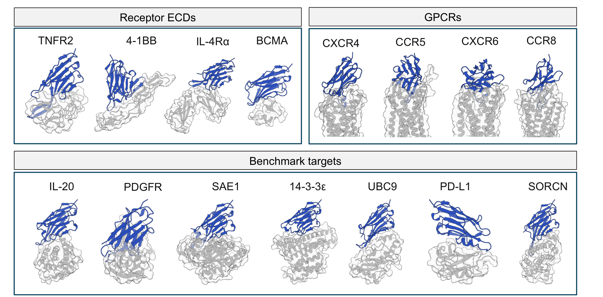
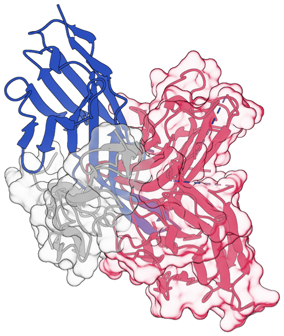
Where a molecule binds can matter as much as whether it binds. For TNFR2 and IL-4Rα, we aimed at the regions used by their natural ligands. The designed VHHs competed with those ligands for binding (Figure 1). These results support binding to the intended ligand-binding regions, highlighting the potential of de novo VHH design to target chosen epitopes. By building that specificity into the design process, we could streamline therapeutic binder discovery and reduce the need for downstream epitope screening.
Building molecules worth taking forward
An exciting binding result becomes more valuable when the molecule also has favorable physical properties. We looked across hydrophobicity, thermal stability, self-interaction, and nonspecific binding to see how often those qualities came together.
Across 30 fully profiled candidates, 23 met at least three of the four criteria. Those candidates covered 12 of 13 targets (Figure 2). Favorable properties appeared across a broad set of designs, giving us more starting points to carry into the next stage of molecular engineering.
From high-affinity binding to cellular activity
4-1BB: compact molecules with potent agonist activity
4-1BB (CD137) helps T cells activate, multiply, and survive, making it an attractive target for cancer immunotherapy (Singh et al., 2024). Tumor-targeted and bispecific 4-1BB agonists offer a way to bring this immune-stimulating activity into multifunctional therapeutics (Klein et al., 2024).
For our 4-1BB designs, we wanted binding to translate into receptor activation. Two VHHs did exactly that: both showed greater agonist potency than Utomilumab in the reporter readout, with activation EC50 values of 0.05 and 0.22 nM, compared with 0.43 nM for Utomilumab (Figure 3).
The appeal extends beyond potency. These designs also combined high purity, thermal stability, and favorable molecular properties. Together, those features make them attractive starting points for building compact 4-1BB modules into bispecific and multispecific antibodies.
| Candidate | Activation EC50 (nM) | SEC purity (280 nm, %) | HIC RT (min) | DSF Tm1 (°C) | AC-SINS (nm) |
|---|---|---|---|---|---|
| 4-1BB VHH 01 | 0.05 | 100 | 7.44 | 70.5 | 0 |
| 4-1BB VHH 02 | 0.22 | 97 | 6.23 | 70.2 | 1 |
One round of feedback changed the next round of design
The first 4-1BB campaign produced two functional hits from 30 designs. We brought those experimental results back into the design process. In the next round, 51 of 60 designs were functional hits—a rise from 7% to 85% (Figure 4).
The next round delivered a much larger pool of potent agonists. This is the part of the story we find especially compelling. Experiments help shape the next set of molecules, turning individual successes into a broader starting point for further molecular engineering.
BCMA: reaching 43 pM affinity from the first design round
BCMA is an established therapeutic target in multiple myeloma, with T-cell engagers and CAR-T therapies already putting it to work. Binding affinity is an important design parameter for BCMA-directed T-cell engagers (Chen et al., 2021). Cilta-cel uses two BCMA-binding VHH domains, illustrating the therapeutic value of compact, single-domain recognition modules (Banerjee et al., 2022).
BCMA offered another striking result. From 30 zero-shot designs, Mol-1 yielded 12 binding hits. One VHH reached an affinity of 43 pM; a second reached 1.1 nM (Figure 5). Here, a computational starting point led directly to exceptionally tight binding.
Both molecules also recognized BCMA on cells. Their cell-binding EC50 values were approximately 22- and 26-fold lower than the belantamab biosimilar comparator. Strong recognition in a compact format provides useful building blocks for multivalent and multispecific antibody designs.
CXCR6: taking design to a membrane receptor
We also took the workflow to CXCR6, a seven-transmembrane chemokine receptor. The designed VHH recognized CXCR6 on cells and antagonized signaling induced by its ligand, CXCL16 (Figure 6). A designed molecule had reached beyond target recognition to change a cellular response.
One VHH, two antigens—by design
Recognizing two antigens with a single VHH is an appealing capability, but finding such molecules through conventional screening can be challenging. We made cross-reactivity a design objective from the outset, asking Mol-1 to create VHHs that recognize both cytokine-A and cytokine-B—two non-orthologous protein antigens sharing only 54% sequence identity.
Five of just 30 designs bound both antigens (Figure 7). To our knowledge, this is the first experimental demonstration of de novo-designed VHHs that cross-react with two non-orthologous protein antigens as intended.
These results show that dual-antigen recognition can be built into the design goal, offering a route to discovering cross-reactive binders with less downstream screening.
Building the next generation of medicines
These results show how biomolecular intelligence can translate into molecules with useful properties—from precise recognition and potent binding to cellular activity. They are an early expression of our broader ambition: to make AI a driving force in the discovery and development of new medicines.
As an AI-native biotech, we bring frontier AI, molecular engineering, and experimental science together to pursue targets and therapeutic possibilities that have remained out of reach. With Mol-1, we aim to push the frontiers of biomolecular intelligence for the benefit of all, bringing us closer to a future where every disease can be cured.
References
- Singh, R., Kim, Y.-H., Lee, S.-J., Eom, H.-S., & Choi, B. K. (2024). 4-1BB immunotherapy: advances and hurdles. Experimental & Molecular Medicine, 56, 32–39. https://doi.org/10.1038/s12276-023-01136-4
- Klein, C., Brinkmann, U., Reichert, J. M., & Kontermann, R. E. (2024). The present and future of bispecific antibodies for cancer therapy. Nature Reviews Drug Discovery, 23, 301–319. https://doi.org/10.1038/s41573-024-00896-6
- Chen, W., Yang, F., Wang, C., Narula, J., Pascua, E., Ni, I., Ding, S., Deng, X., Chu, M. L.-H., Pham, A., Jiang, X., Lindquist, K. C., Doonan, P. J., Van Blarcom, T., Yeung, Y. A., & Chaparro-Riggers, J. (2021). One size does not fit all: navigating the multi-dimensional space to optimize T-cell engaging protein therapeutics. mAbs, 13, 1871171. https://doi.org/10.1080/19420862.2020.1871171
- Banerjee, R., Lee, S. S., & Cowan, A. J. (2022). Innovation in BCMA CAR-T therapy: Building beyond the Model T. Frontiers in Oncology, 12, 1070353. https://doi.org/10.3389/fonc.2022.1070353